Intra-MRI Head Motion Tracking and Correction: A Quantitative In Vivo Evaluation Framework
Summary
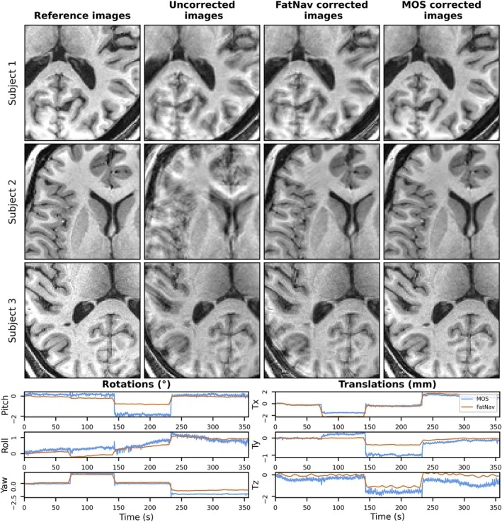
Volunteers made visually guided head rotations during MRI, allowing the researchers to compare optical tracking and MR-based fat navigators against a reference estimate. Optical tracking gave better corrected images overall. The reference measurements also revealed direction-dependent differences in tracking precision.